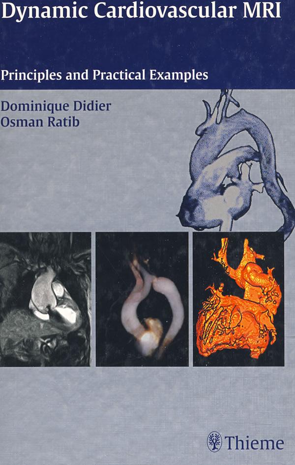
动力心血管MRI Dynamic Cardiovascular MRI
 点此进入淘宝搜索页搜索
分类: 图书,进口原版书,医学 Medicine ,
作者: Dominique Didier, Osman Ratib著
出 版 社: Oversea Publishing House
出版时间: 2003-4-1字数:版次: 1页数: 186印刷时间:开本: 16开印次: 1纸张:I S B N : 9783131330413包装: 精装内容简介
Recent progress in MR imaging techniques has lead to a rapid increase in the number of clinical applications that benefit from the non—invasive imaging Of cardiovascular structures.These
innovative imaging techniques present US with unique abilities for the investigation of anatomical structures as well as the functional performance of the heart and thoracic great vessels.
This book and companion CD richly illustrate—with carefully selected pictures and dynamic video of typical clinical cases—the basic principles 0f cardiovascular MR imaging techniques.while also providing a comprehensive review of the clinical applications of these techniques.The book iS conveniently organized into seven main chapters covering congenital heart disease,aortic anomalies,cardiac masses,valvular diseases。pericardial diseases,cardiomyo—pathies and ischemic heart disease.
目录
1 Cardiac MR Imaging Techniques
1.1 Standard Views in Cardiac Imaging
1.1.1 Orthogonal Image Plane Orientation
1.1.2 Oblique Plane Positioning
1.1.3 Standard Orthogonal Views of the Heart
1.1.4 Oblique Cardiac Imaging Planes
1.2 Techniques and Imaging Sequences
1.2.1 Cardiac Gating(ECG Gating)
1.2.2 Lead Placement
1.2.3 How to Improve the Trace
1.2.4 MR Imaging Sequences
1.2.5 Multiplanar Scout Images(Localizers)
1.2.6 Cardiovascular Morphology
1.2.7 Cardiac Function
1.2.8 MR Angiography(MRA)Techniques
1.2.9 Contrast—Enhanced MRA
1.2.10 Flow Quantification and Velocity—Encoded Cine Sequences
1.3 Post—Processing and Rendering Techniques
1.3.1 Multiplanar Reformatting(MPR)
1.3.2 “Thick—Slab”MPR
1.3.3 Curved—Oblique Reformatted Images
1.3.4 Maximum Intensity Projections(MIP)
1.3.5 Surface Rendering and Volume Rendering
2Congenital Heart Disease
2.1 Major Indications of MRlin CHD
2.2 Imaging Techniques
2.2.1 Sedation.Monitoring and Patient Preparation
2.2.2 Spin—Echo Technique
2.2.3 Cine—MR Imaging
2.2.4 Flow—Sensitive Imaging Techniques
2.2.5 Gadolinium—Enhanced 3D MR Angiography
2.3 Segmental Description of Cardiac Anomalies
2.4 Thoracic Aortic and Aortic Arch Anomalies
2.5 Cono-Truncal MalfcIrmations and Complex Anomalies
2.5.1 Tetralogy of Fallot
2.5.2 Pulmonary Atresia
2.5.3 Double Outlet Right Ventricle
2.5.4 Truncus Arteriosus
2.5.5 Transposition of the Great Arteries
2.5.6 Single Ventricle
2.5.7 Tricuspid Atresia
2.6 Pulmonary and Systemic Venous Anomalies
2.7 Detection and Quantitative Analysis of Shunts.Stenoses and Regurgitations
2.8 Other Miscellaneous Anomalies
2.8.1 Ebstein’S Anomaly
2.8.2 Cor Triatriatum
2.9 Postoperative Evaluation of CHD in Adult Patients
2.9.1 Patency and Complications in Systemic—to—Pulmonary Artery Shunts
2.9.2 Surgical Repair of Tetralogy of Fallot and Cono-Truncal Malformations
2.9.3 Intracardiac Baffles and Patches
2.9.4 Arterial Switch Operation for Transposition of the Great Vessels
3 Throacic Aorta
3.1 Coarctation of the Aorta
3.2 Aortic Arch Anomalies
3.3 Interrupted Aortic Arch
3.4 Cervical Aortic Arch
3.5 Aortic Aneurvsms
3.6 Aortic Dissections
3.7 AOrtitis
3.8 Aortic Thrombus
4 Cardiac Masses
4.1 ClassificatiOn and Characte rization of Cardiac Masses
4.2 Primary Cardiac Tumors—Benign Tumors
4.2.1 Myxoma
4.2.2 Lipoma and Lipomatous Atrial Septum
4.2.3 Fibroma
4.2.4 Rhabdomyomas
4.3 Primary Cardiac Tumors—Malignant Tumors
4.3.1 Angiosarcoma
4.3.2 Rhabdomyosarcoma and Fibrosarcoma
4.3.3 Mesothelioma
4.3.4 Malignant Fibrous Histiocytoma
4.4 Secondary Cardiac Tumors
4.5 Non-Tumoral Cardiac Masses
……
5 Valvular Heart Disease
6 Pericardial Disease and Cardiomyopathy
7 Ischemic Heart Disease
8 Index
9 Dynamic Cardiac MRI-Companion CD